

Conditions we treat
We treat both medical and surgical diseases of the retina, macula and vitreous.

Macular Degeneration
This disease is characterized by the deterioration of the central portion of the retina, which is responsible for recording the images we see and sending them via the optic nerve from the eye to the brain. The retina’s central portion – the macula – is responsible for focusing central vision in the eye, and it controls our ability to read, drive a car, recognize faces or colors, and see objects in fine detail.
“This image was originally published in the Retina Image Bank. Mallika Goyal, MD. Advanced Wet AMD. Retina Image Bank. 2013; 12163. © the American Society of Retina Specialists."

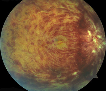
Retinal Tears and Detachments
When the retina tears and pulls away from its normal position at the back of the eye, the retina is considered to be “detached.” Once pulled away, the retina no longer works properly and loses its blood supply. Retinal tears and detachments are serious problems that result in blindness unless treated.
“This image was originally published in the Retina Image Bank. Homayoun Tabandeh, MD, FASRS. Retinal Tear at the Posterior Edge of Lattice Degeneration. Retina Image Bank. 2014; 14662. © the American Society of Retina Specialists."

Diabetic Retinopathy
If you have diabetes mellitus, your body does not use and store sugar properly.High blood sugar levels cause damage to the small blood vessels in the retina. The damage to retinal vessels is called diabetic retinopathy.
“This image was originally published in the Retina Image Bank. Theodore Leng, MD, MS. Proliferative Diabetic Retinopathy. Retina Image Bank. 2013. © the American Society of Retina Specialists."

Macular Holes
Macular holes can form when the vitreous, the gel-like substance that fills the eye and lies in front of the macula, shrinks and pulls away from the macula. In some instances, the vitreous gel sticks to the macula and cannot pull away, stretching the macular tissue, and ultimately tearing.
“This image was originally published in the Retina Image Bank. Yusuke Oshima, MD, PhD. Yusuke Takada, Osaka University Graduate School of Medicine. Idiopathic Macular Hole. Retina Image Bank. 2013; 5257. © the American Society of Retina Specialists."

Flashes and Floaters
Floaters look like small specks, dots, circles, lines or cobwebs in your field of vision, while flashes can look like flashing lights or lightning streaks in your field of vision.
“This image was originally published in the Retina Image Bank. Henry J. Kaplan, MD. Asteroid Hyalosis. Retina Image Bank. 2013; 5455. © the American Society of Retina Specialists."

Macular Edema
With this condition, fluid leaks from retinal blood vessels, causing swelling or thickening of the macula. This is called macular edema and results in blurring of the central vision.
“This image was originally published in the Retina Image Bank. Jason S. Calhoun, Department of Ophthalmology, Mayo Clinic Jacksonville, Florida. Cystoid Macular Edema. Retina Image Bank. 2013; 7670. © the American Society of Retina Specialists."
Retinal Vein Occlusion
A retinal vein occlusion occurs when a vein in the retina of the eye is blocked. A blocked vein damages the blood vessels of the retina, causing bleeding and leakage of fluid from the areas of blocked blood vessels.
“This image was originally published in the Retina Image Bank. Lihteh Wu, MD. Central Retinal Vein Occlusion. Retina Image Bank. 2012; 1887. © the American Society of Retina Specialists."